
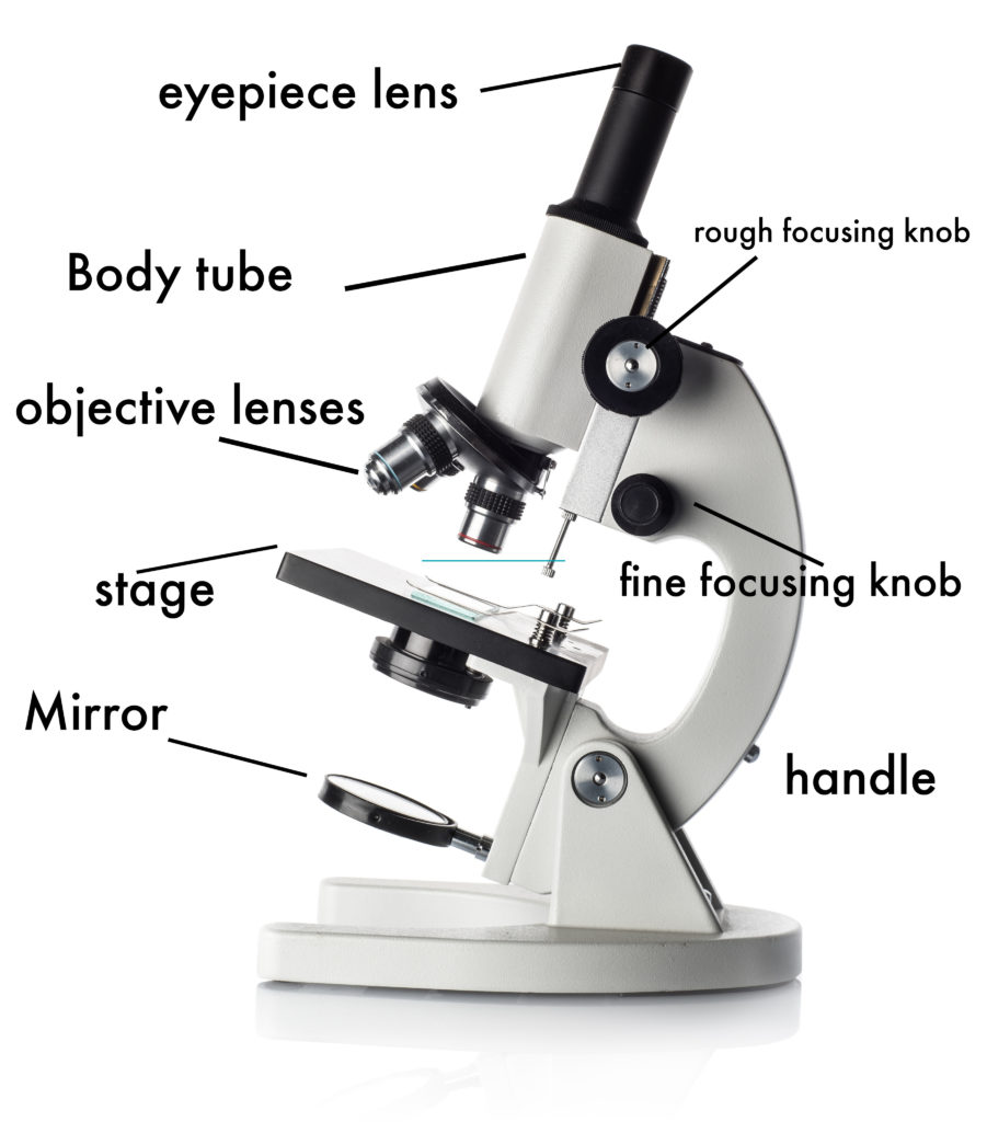
Draw And Label Microscope - Eyepiece (10x) and objective lenses (4x, 10x, 40x, 100x).
Draw And Label Microscope - Web ready to take your drawing skills to the next level? In this interactive, you can label the different parts of a microscope. Web download the label the parts of the microscope pdf printable version here. Web use this interactive to identify and label the main parts of a microscope. There are six printables available.
Draw the objective lenses 1.5 step 5: The part that is looked through at the top of the compound microscope. Continue follow my channel and like, share,comm. Draw the base of the microscope sketch 1.7 step 7: Perfect for students or anyone. The lens the viewer looks through to see the specimen. Microscope world explains the parts of the microscope, including a printable worksheet for.

Microscope Diagram Labeled, Unlabeled and Blank Parts of a Microscope
Web common compound microscope parts include: Also indicate the estimated cell size in micrometers under your drawing. The lens the viewer looks through to see the specimen. Review the principles of light microscopy and identify the major parts of the microscope. Microscope world explains the parts of the microscope, including a printable worksheet for. Shape.
The Wonders Of Microscopes What You Need To Know Creyentes Diverses News
Eyepiece (10x) and objective lenses (4x, 10x, 40x, 100x). First and foremost, we have a labeled microscope diagram, available in both black and white and color. Describe the functions of each part of the microscope you have drawn above. Web how to draw a microscope 🔬. Provide them with diagrams or actual microscopes, and guide.

Parts of a microscope with functions and labeled diagram
It is used to observe things that cannot be seen by the naked eye. Learn how to use the microscope to view slides of several different cell types, including the use of the oil immersion lens to view bacterial cells. The lens the viewer looks through to see the specimen. Web there are three major.

Microscope diagram Tom Butler Science skills, Microscope parts
Draw the objective lenses 1.5 step 5: Web art for kids hub. Microscope world explains the parts of the microscope, including a printable worksheet for. Draw the base of the microscope sketch 1.7 step 7: Useful as a means to change focus on one eyepiece so as to correct for any difference in vision between.

Compound Microscope Parts Labeled Diagram and their Functions (2023)
Differentiate between a condenser and an abbe condenser. Provide them with diagrams or actual microscopes, and guide them through the function of each part, such as the lens, eyepiece, and stage. Web common compound microscope parts include: Web a microscope is one of the invaluable tools in the laboratory setting. The body tube connects the.

Microscope Drawing And Label at GetDrawings Free download
Outline the slide platform 1.6 step 6: Outline the arm frame 1.4 step 4: Web ready to take your drawing skills to the next level? Download the label the parts of the microscope: Answers pdf printable version here. Download the diagrams and practice labeling the different parts of these. Learn how to use the microscope.

Compound Light Microscope Drawing at GetDrawings Free download
Useful as a study guide for learning the anatomy of a microscope. And drop the text labels onto the microscope diagram. Web ready to take your drawing skills to the next level? Outline the slide platform 1.6 step 6: Perfect for students or anyone. Always lift a microscope by holding both the arm and base.

Label the Microscope Diagram Download Scientific Diagram
Differentiate between a condenser and an abbe condenser. We’ll have covered the parts of both simple and compound microscopes and their functions in this article. Web table of contents 1 how to draw a microscope that is hyperrealistic 1.1 step 1: Most photographs of cells are taken using a microscope, and these pictures can also.

5 Types of Microscopes with Definitions, Principle, Uses, Labeled Diagrams
Knobs (fine and coarse) 6. Web art for kids hub. There are six printables available. Microscope world explains the parts of the microscope, including a printable worksheet for. Web ready to take your drawing skills to the next level? The part that is looked through at the top of the compound microscope. Outline the arm.

36+ Label Each Part Of A Microscope Gif Diagram Printabel
The working principle of a simple microscope is that when a lens is held close to the eye, a virtual, magnified and erect image of a specimen is formed at the least possible distance from which a human. In this tutorial, writing master shows you how to draw a realistic microscope with labels step by.
Draw And Label Microscope Useful as a study guide for learning the anatomy of a microscope. Web pencil drawing paper crayons or colored pencils black marker (optional) draw a microscope printable pdf (see bottom of lesson) the goal is to complete a drawing of a microscope by creating each part one part at a time. Describe the functions of each part of the microscope you have drawn above. Diagrammatically, identify the various parts of a microscope. The lens the viewer looks through to see the specimen.
First And Foremost, We Have A Labeled Microscope Diagram, Available In Both Black And White And Color.
Perfect for students or anyone. Microscope world explains the parts of the microscope, including a printable worksheet for. Label the cell wall, cell membrane, cytoplasm, and chloroplasts in your lab manual. Also indicate the estimated cell size in micrometers under your drawing.
The Lens The Viewer Looks Through To See The Specimen.
Answers pdf printable version here. Useful as a study guide for learning the anatomy of a microscope. Download the diagrams and practice labeling the different parts of these. Differentiate between a condenser and an abbe condenser.
Web Use This Interactive To Identify And Label The Main Parts Of A Microscope.
Eyepiece (10x) and objective lenses (4x, 10x, 40x, 100x). Web there are three major structural parts of a microscope: The body tube connects the eyepiece to the objective lenses. Today, we're learning how to draw a cool microscope!👩🎨 join our art hub membership!
Outline The Arm Frame 1.4 Step 4:
Web table of contents 1 how to draw a microscope that is hyperrealistic 1.1 step 1: Diagrammatically, identify the various parts of a microscope. Knobs (fine and coarse) 6. There are two major optical lens parts of a microscope: